Tin tức
A STEP-BY-STEP VISUAL STORY THAT TAKES THE VIEWER THROUGH THE PATHOLOGY LAB PROCESS.
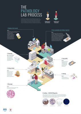

Booking in and labelling specimen by laboratory personnel
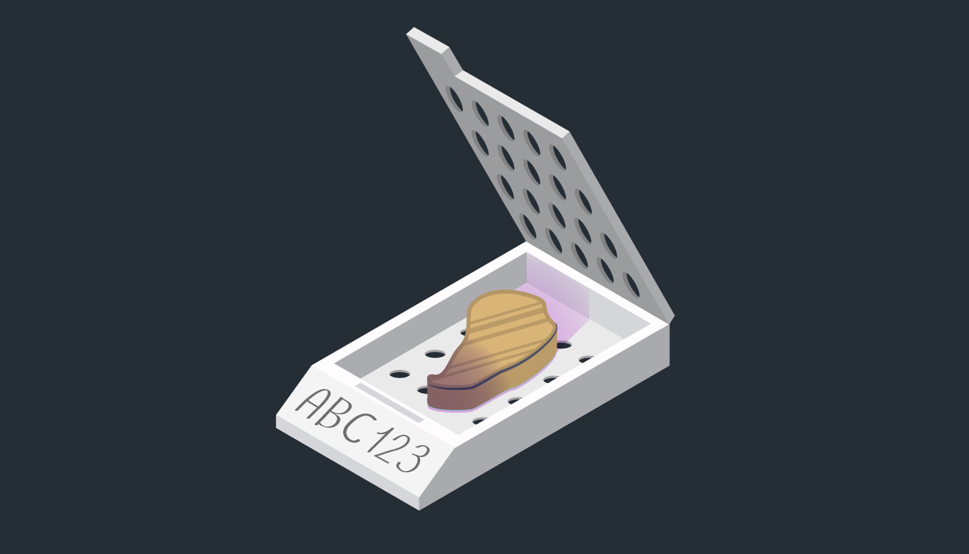
Cut specimens are placed in small, labelled containers called cassettes.

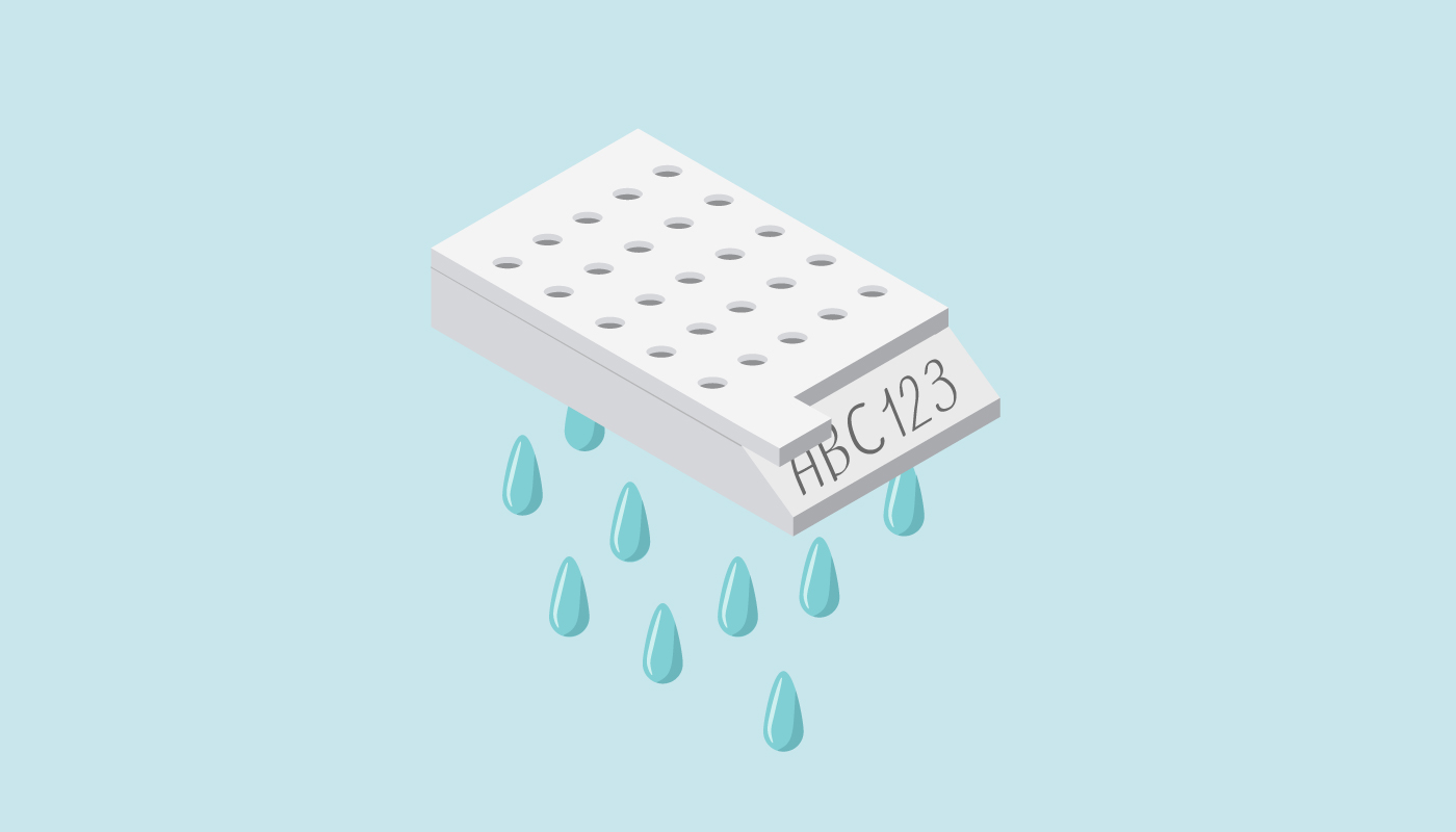
Specimen container with barcode for identification
Gross examination, recording and cutting of specimen carried out by pathologist
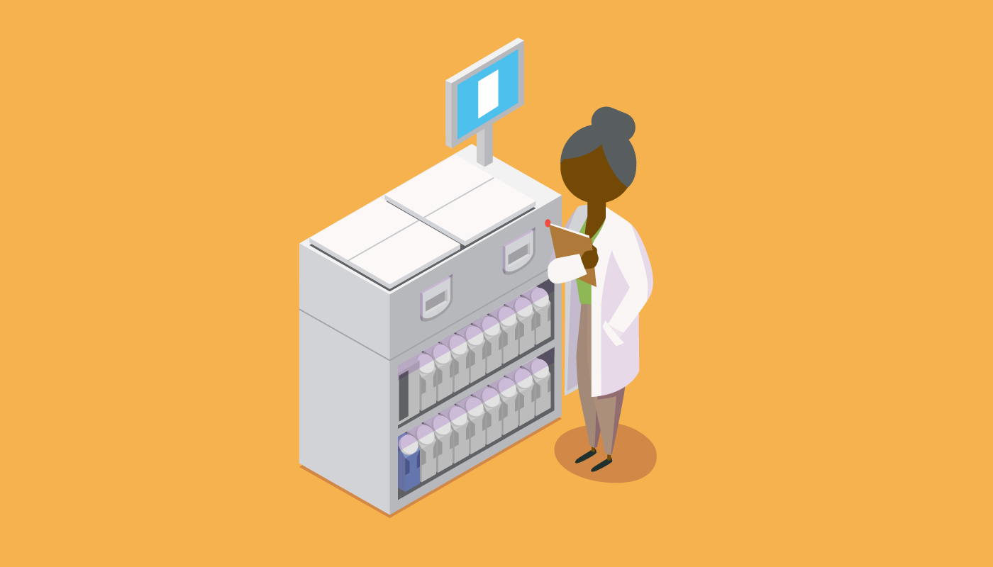
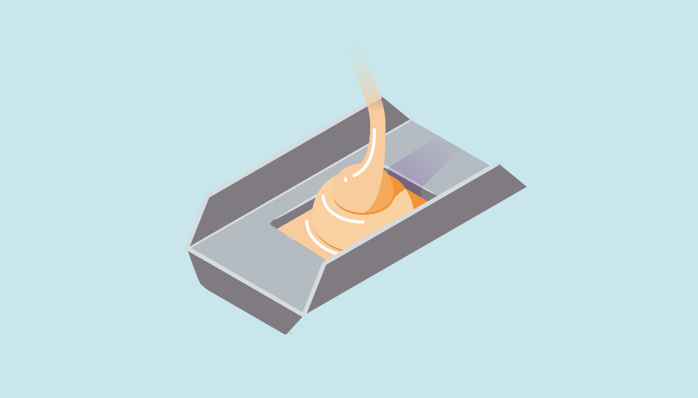
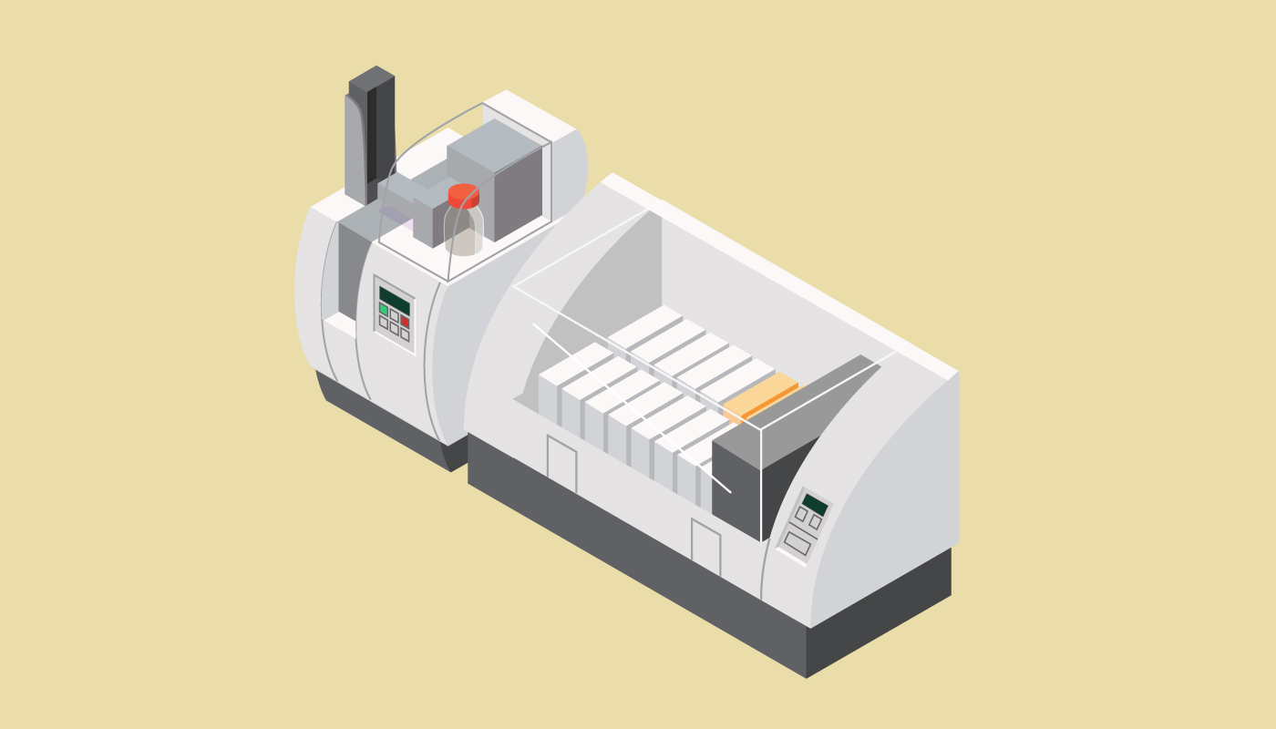
Cassettes are placed into a processor overnight
Processor gradually removes water from the specimen
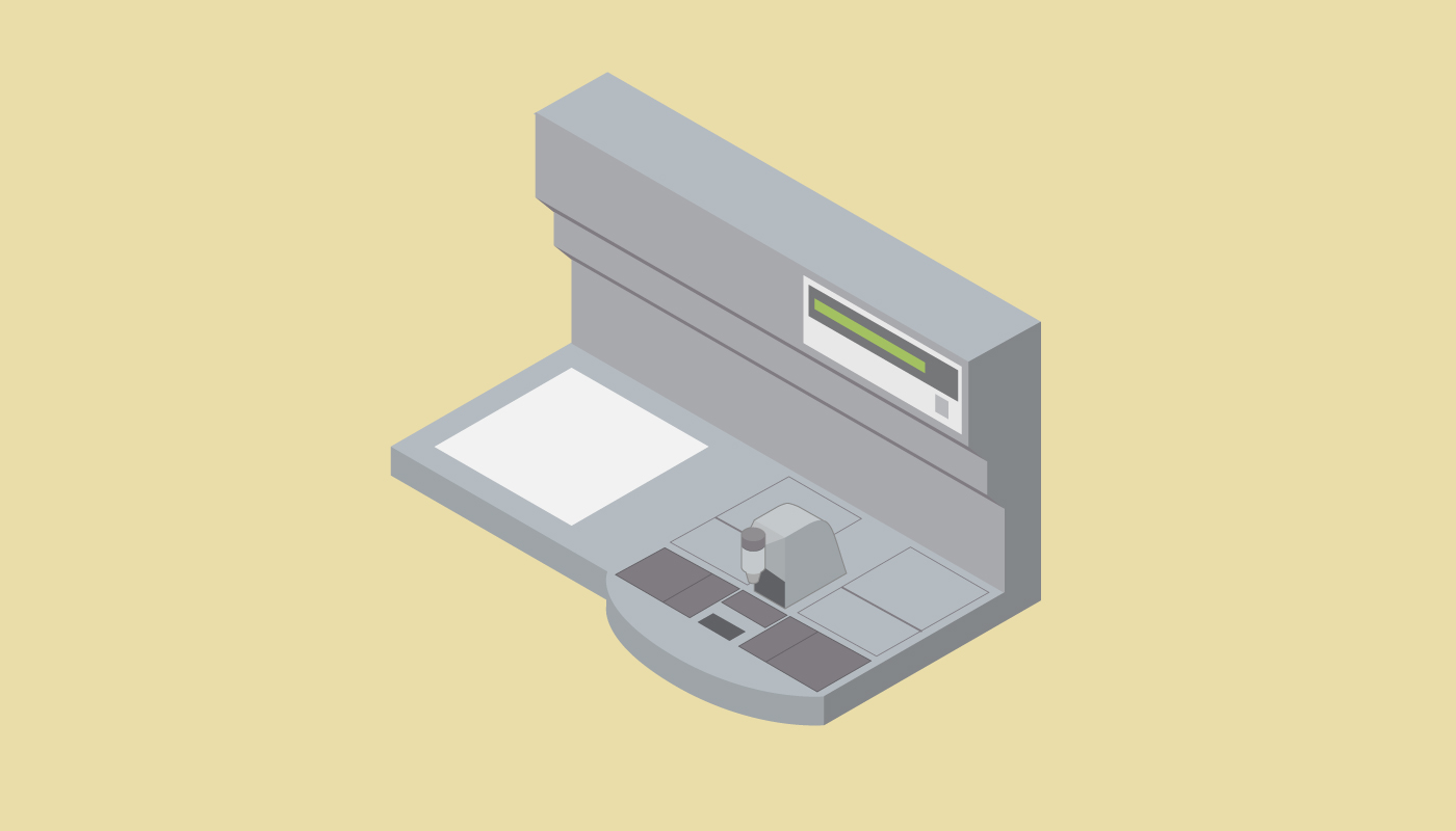
Processed tissue fragments are taken out of the processor and cassettes and transferred to a tissue embedding station.
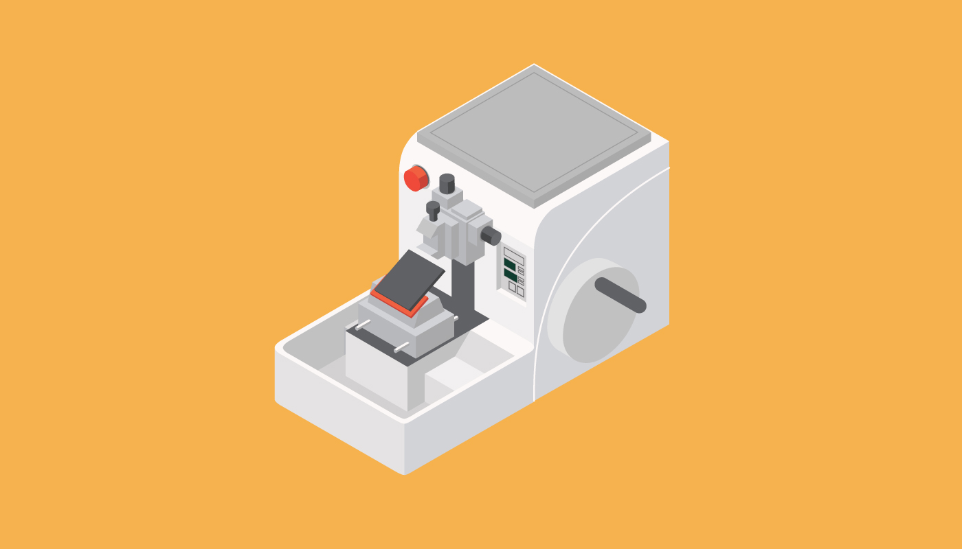
Wax blocks are then transferred to a microtome station.
Specimen segments are then embedded in paraffin wax blocks that harden at room temperature.

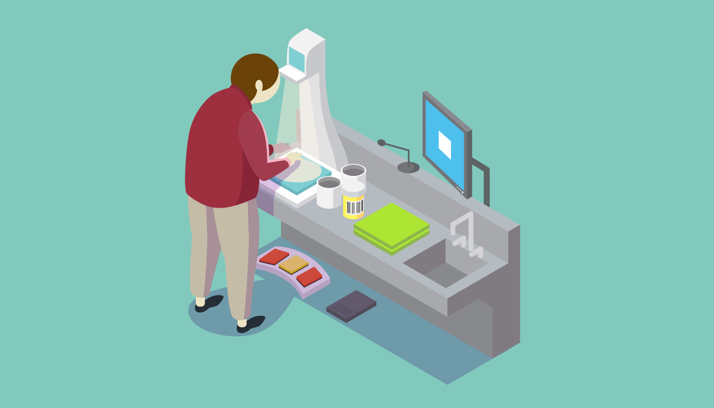
The microtome cuts the wax blocks into extremely thin slices.
Thin specimen slices are placed on a slide and put through an automatic slide coverslipper before being put through a stainer.
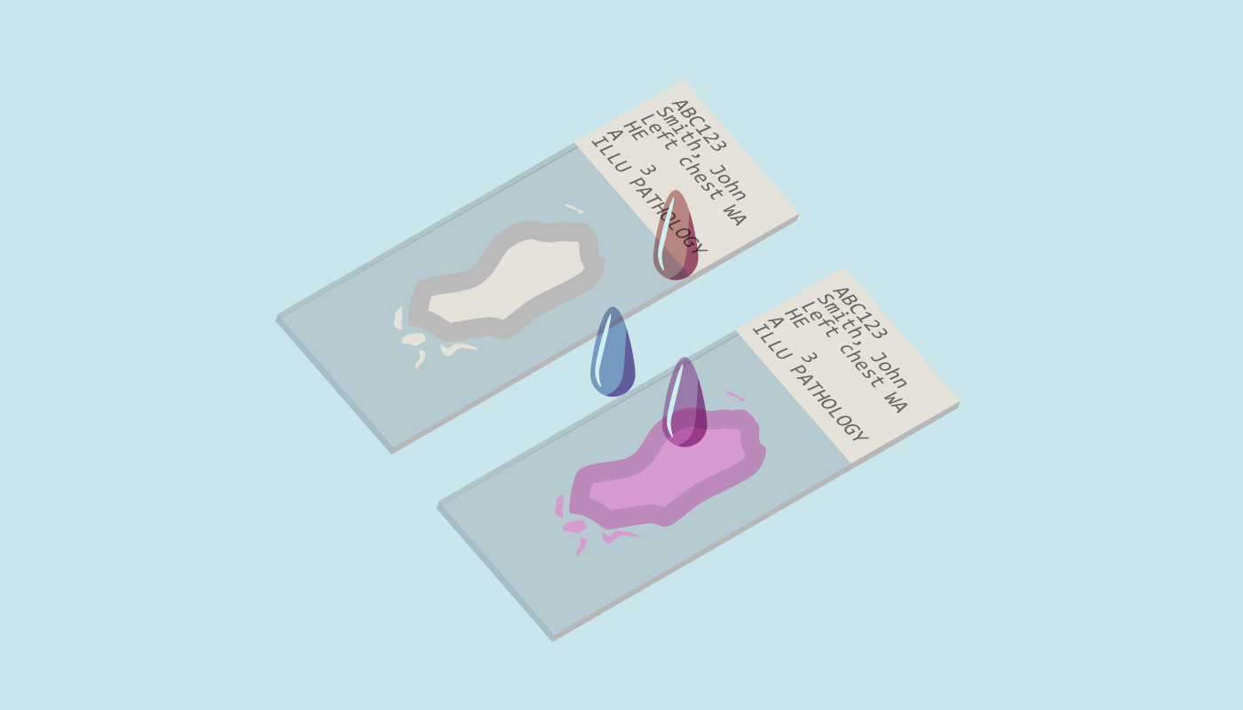
The specimen slides are stained with eosin (red) and haematoxylin (blue).
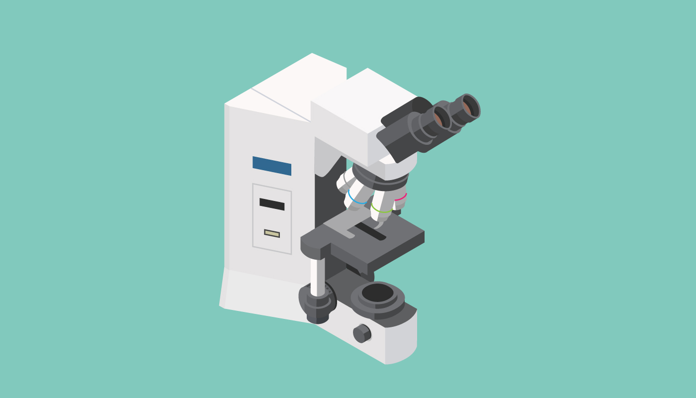
Once stained the slides are then ready for review by the pathologist through a microscope. They will then be able to make a diagnosis or undertake further techniques to aid in diagnosis.